Research

Bacteria make up the majority of life on Earth. They can be found everywhere: ocean, air, soil, animals, plants, etc. Their impact is monumental. For instance:
– in the soil, bacteria are stakeholders in the cycle of regeneration of agricultural land
– in the health, they are essential for the wellness of humans (intestinal flora, microbiota, skin barrier, etc)
– in the industry, they are used as a machine to synthesize drugs (insulin, antibiotics, etc.)
– in the medecine, they cause diseases
– on the environment, they incur the eutrophication of the oceans
– …

However, we are still scratching the surface of the bacteria significance. Some refer to them as the “dark matter alive”.

Major questions remain, such as:
– What kind of bacteria are living on Earth? (most of them do not grow in the laboratory of scientists to be easily indentify)
– What are the interplay between the bacteria and  their environment?
– How do they interact with their neighbourhood?
– Do they communicate? If yes, how?
– Does bacterial population organize themselves?
– How do they adapt to their changing environment?

Thus, the stage is set for many important features to be explored and discovered.
The following is presenting research projects aiming at progressing toward the answer of these essential questions. Feel free to scroll down and have a look.

Biofilms and cellulose – biophysical consequences

See our paper on biofilms and the pathway controlling the cellulose production at the air-liquid-interface.

Title: Causes and biophysical consequences of cellulose production by Pseudomonas fluorescens SBW25 at the air-liquid interface
Authors: Maxime Ardré, Djinthana Dufour, Paul B Rainey

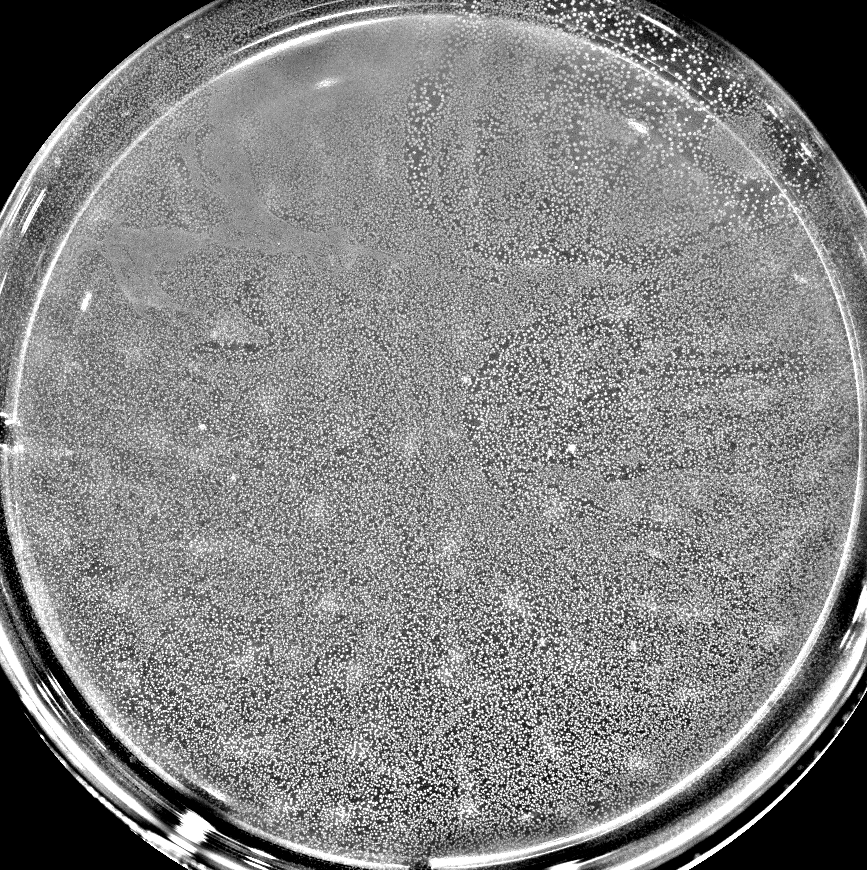
Biofilm formation by P. fluorescens at the air-liquid interface (ALI). The photo is a top view of a well (40mm diameter).
The bacteria produce small white dots on the interface by up regulating the cellulose production locally. The more white areas scattered on the surface indicate the presence of active convection into the liquid below. This convection is driven by the biomass increase at the ALI.
We decipher the importance of several metabolic pathways involved in the cellulose production. We give a mathematical model that explains the apparition of streams. We rationalize the consequences of the streams over the spatial distribution of extracellular products produced by the bacteria within the biofilm.

Mechanics of microcolonies

“Surface colonization underpins microbial ecology on terrestrial environments. Although bacterial adhesion has been extensively studied, its dynamical contribution to colony expansion remains largely unexplored. Here, we used laser ablation and force microscopy to monitor adhesion at the single-cell level. We show that adhesion forces of rod-shaped bacteria are higher at old poles. This asymmetry induces mechanical tension, and drives daughter cell rearrangements, which eventually determine colony shape. Informed by microscopic data, we developed a quantitative model for colony morphogenesis that fits experiments, and predicts bacterial adhesion from simple time-lapse measurements. Our results demonstrate that patterns of surface colonization are shaped by force relaxation events derived from an asymmetric distribution of adhesive factors at cell poles.”

Title: Asymmetric adhesion of rod-shaped bacteria controls microcolony morphogenesis

Authors: Duvernoy Marie-Cécilia, Mora Thierry, Ardré Maxime, Croquette Vincent, Bensimon David, Quilliet Catherine, Ghigo Jean-Marc, Balland Martial, Beloin Christophe, Lecuyer Sigolène, Desprat Nicolas

Bacteria in droplets

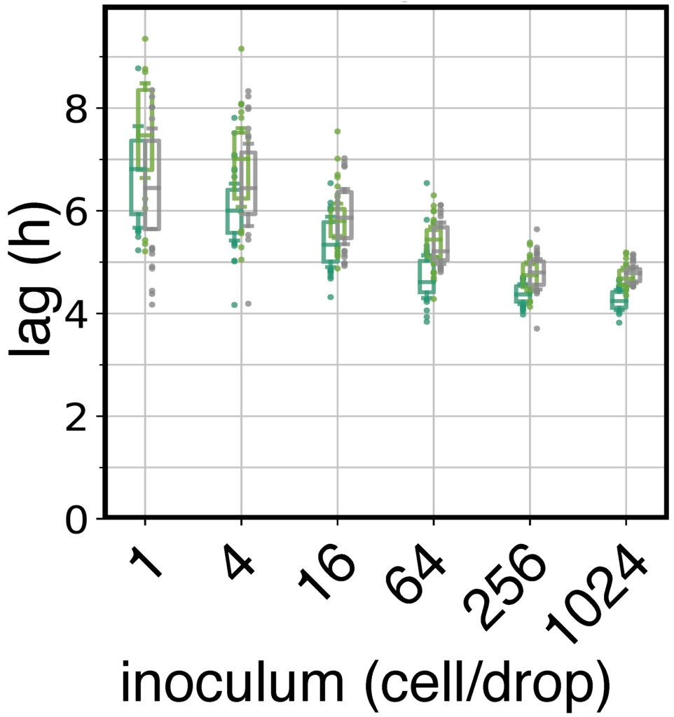
“The relationship between the number of cells colonizing a new environment and time for resumption of growth is a subject of long-standing interest. In microbiology this is known as the “inoculum effect.” Its mechanistic basis is unclear with possible explanations ranging from the independent actions of individual cells, to collective actions of populations of cells. Here, we use a millifluidic droplet device in which the growth dynamics of hundreds of populations founded by controlled numbers of Pseudomonas fluorescens cells, ranging from a single cell, to one thousand cells, were followed in real time. Our data show that lag phase decreases with inoculum size. The decrease of average lag time and its variance across droplets, as well as lag time distribution shapes, follow predictions of extreme value theory, where the inoculum lag time is determined by the minimum value sampled from the single-cell distribution. Our  experimental results show that exit from lag phase depends on strong interactions among cells, consistent with a “leader cell” triggering end of lag phase for the entire population.”

Title: A leader cell triggers end of lag phase in populations of Pseudomonas fluorescens

Authors : Maxime Ardré, Guilhem Doulcier, Naama Brenner, Paul B Rainey

Modelling of Biofilm formation dynamics

“The bacterium Bacillus subtilis frequently forms biofilms at the interface between the culture medium and the air. We present a mathematical model that couples a description of bacteria as individual discrete objects to the standard advection-diffusion equations for the environment. The model takes into account two different bacterial phenotypes. In the motile state, bacteria swim and perform a run- and-tumble motion that is biased toward regions of high oxygen concentration (aerotaxis). In the matrix-producer state they excrete extracellular polymers, which allows them to connect to other bacteria and to form a biofilm. Bacteria are also advected by thefluid, and can trigger bioconvection. Numerical simulations of the model reproduce all the stages of biofilmformation observed in laboratory experiments. Finally, we study the influence of various model parameters on the dynamics and morphology of biofilms.”

Title: An individual-based model for biofilm formation at liquid surfaces

Authors: Maxime Ardré, Hervé Henry , Carine Douarche and Mathis Plapp

Microfluidic

A first approach is to monitor the bacteria at the single cell level. To do so I developed a microfluidic chip integrated to a microscope in order to control the chemical environment of the bacteria. The microfluidic technology sketched in the image above allows to keep a steady environment at the bacterial scale. Thanks to this device we can follow the growth of microcolonies such as in the movie presented here.

From this movie big data analysis (neuronal network) can be performed to segment the images and extract valuable informations from the dynamical system.

The top part of the movie shows the bacteria producing an important molecules. The bottom part of the movie shows the shape of the same bacteria. We can see that the rod shape bacteria grow and split themselves with time to form microcolonies (few micrometers).  In the same time the production of the important molecule is specific to each bacteria and “shared” between neighboors.